Our X-ray Melbourne Statements
Table of ContentsFascination About Bulk-billed Ct ScansCat Scan Melbourne Fundamentals ExplainedThe Buzz on Bulk-billed Ct Scans
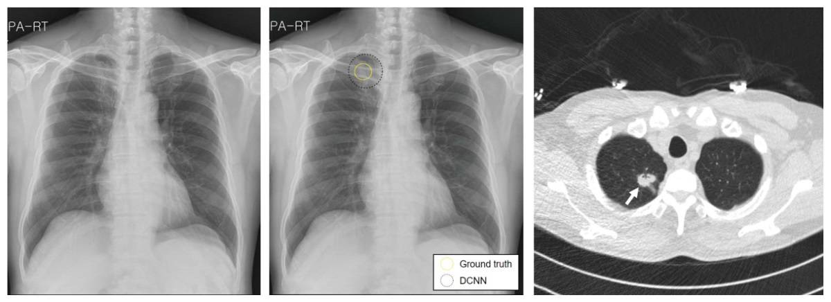
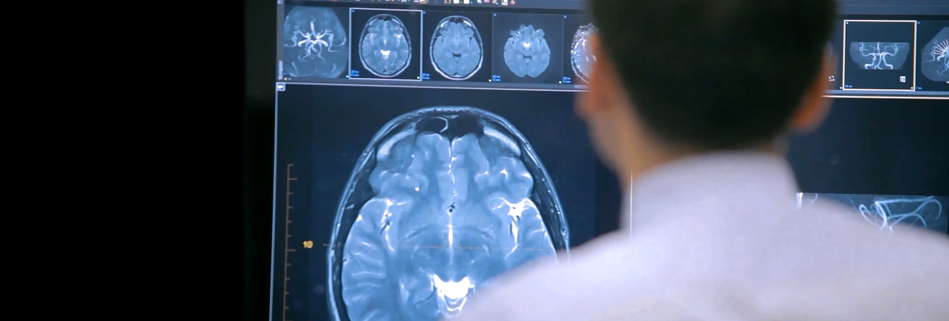
Dr. Macintyre's X-Ray Film (1896) Radiology is the clinical technique that uses medical imaging to diagnose and also deal with conditions within the bodies of pets, including human beings. A range of imaging methods such as X-ray radiography, ultrasound, computed tomography (CT), nuclear medicine consisting of positron emission tomography (ANIMAL), as well as magnetic vibration imaging (MRI) are made use of to identify or deal with diseases.The modern-day technique of radiology entails numerous various medical care careers functioning as a group. The radiologist is a clinical doctor that has actually completed the ideal post-graduate training as well as analyzes medical images, communicates these findings to various other medical professionals using a record or verbally, and makes use of imaging to do minimally invasive clinical treatments.

The X-rays are forecasted via the body onto a detector; a picture is formed based on which rays travel through (as well as are spotted) versus those that are taken in or spread in the individual (and therefore are not detected). Rntgen uncovered X-rays on November 8, 1895 and obtained the first Nobel Prize in Physics for their exploration in 1901.
The X-rays that travel through the patient are filteringed system through a device called an grid or X-ray filter, to lower scatter, and also strike an undeveloped movie, which is held tightly to a screen of light-emitting phosphors in a light-tight cassette. The movie is then created chemically as well as a picture appears on the film.
In the 2 latest systems, the X-rays strike sensing units that transforms the signals generated into digital information, which is transferred and also transformed into a photo displayed on a computer system display. In electronic radiography the sensing units form a plate, however in the EOS system, which is a slot-scanning system, a linear sensor vertically checks the individual.
The 10-Second Trick For Bulk-billed X-rays
Due to its schedule, rate, and also lower prices compared to various other methods, radiography is typically the first-line test of option in radiologic diagnosis. Also despite the huge amount of information in CT scans, MR scans and other digital-based imaging, there are many disease entities in which the traditional diagnosis is obtained by plain radiographs - Cat Scan Melbourne.
Mammography and DXA are two applications of reduced power projectional radiography, made use of for the evaluation for bust cancer as well as osteoporosis, respectively. Fluoroscopy and angiography are unique applications of X-ray imaging, in which a fluorescent display and picture intensifier tube is linked to a closed-circuit television system.:26 This enables real-time imaging of structures moving or enhanced with a radiocontrast agent.
Two radiocontrast agents are presently alike use. Barium sulfate (BaSO4) is provided orally or rectally for evaluation of the GI system. Iodine, in several proprietary kinds, is provided by dental, anal, genital, intra-arterial or intravenous paths. These radiocontrast representatives highly soak up or scatter X-rays, as well as combined with the real-time imaging, enable demonstration of dynamic procedures, such as peristalsis in the digestive system tract or blood circulation in arteries and also veins.


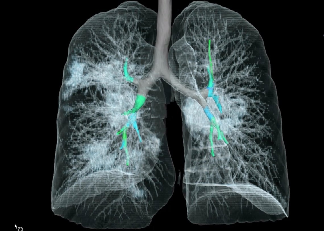
With fast administration of intravenous contrast during the CT scan, these great detail images can be rebuilded right into three-dimensional (3D) pictures of carotid, analytical, coronary or various other arteries. The intro of computed tomography in the very early 1970s transformed analysis radiology by giving Clinicians with photos of real three-dimensional anatomic structures.
The 20-Second Trick For X-ray Melbourne


The initial ultrasound pictures were static and two-dimensional (2D), however with modern-day ultrasonography, 3D reconstructions can be observed in genuine time, successfully ending up being "4D". Because ultrasound imaging techniques do not employ ionizing radiation to generate images (unlike radiography, and also CT scans), they are normally thought about more secure and also are therefore extra typical in obstetrical imaging.

Ultrasounds is valuable as a guide to executing biopsies to reduce damages to bordering cells and in drainages such as thoracentesis. Little, portable ultrasound devices now change peritoneal lavage in trauma wards by non-invasively assessing for the presence of internal bleeding as well as any type of interior body organ damage. Extensive interior blood loss or injury to the major body organs might call for surgery and also repair service. X-Ray Melbourne.